
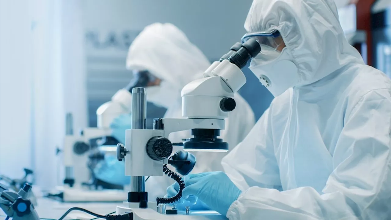
In this interview, News-Medical.net speaks to Prof. Klaus Gerwert of Ruhr University, about how histopathology problems can be solved using infrared microscopy.
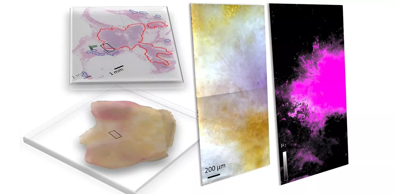
What is histopathology and how is microscopy used in conjunction with histopathology to diagnose disease? In classical histopathology, you take a biopsy, slice the biopsy in thin sections, and then you stain the biopsy with hematoxylin and eosin stain , a specific compound. This visualizes some morphological changes within the tissue. The pathologist then looks at these morphological changes and they can classify the disease and identify if there is cancer present or not.
We have shown by using the quantum cascade laser based microscope that you can do all of these differential diagnoses and that this is what the clinicians need in order to make a specific therapeutic decision. Now we need to look at sensitivity and specificity versus the new label-free digital pathology approach and compare it to the classical histopathology approach, which is, at the moment, the gold standard in the clinics.
Problem Solving Classical Histopathology with IR Imaging MicroscopyPlay Related StoriesUsing bioinformatics, you can assign a specific color to the fingerprint. Then you've got an indexed color image, and this indexed color image is equivalent to the H&E stain picture in classical histopathology. The big advantage of this is that you can always use the same classifier. This eliminates any variability. This is a further big advantage.
As a result, QCL is really a big breakthrough in infrared spectroscopy. In contrast to FTIR, which takes about 20 hours to take a measurement, QCL can take the same measurements in 20 minutes.
Microscopy Adenocarcinoma Biopsy Cancer Compound Diagnostics Digital Pathology Microscope Pathology Protein Spectroscopy
United Kingdom Latest News, United Kingdom Headlines
Similar News:You can also read news stories similar to this one that we have collected from other news sources.
 Innovative microscopy demystifies metabolism of Alzheimer'sAlzheimer's disease causes significant problems with memory, thinking and behavior and is the most common form of dementia, affecting more than 50 million people around the world each year. This number is expected to triple by the year 2050.
Read more »
 Electron Microscopy in Industry: Quality Control and Failure AnalysisElectron microscopy enhances nanoscale analysis for quality control and failure analysis across diverse industries.
Read more »
 Emerging Electron Microscopy Techniques for Quantum ResearchAdvanced electron microscopy unlocks the secrets of quantum materials, enabling high-resolution characterization of their unique quantum properties.
Read more »
 Study introduces hyperspectral dark-field microscopy for rapid and accurate identification of cancerous tissuesBreast-conserving surgery (BCS), also called lumpectomy, involves the removal of a cancerous lump and some surrounding tissue. BCS is suitable for women with early-stage breast cancer or small lumps, as it preserves more of the breast compared to mastectomy.
Read more »
 Recent Advances in High-Speed Atomic Force Microscopy to Improve Research Into Protein DynamicsIn this interview, AZoNano speaks with Dr. George Heath from the University of Leeds, UK, about the fundamental principles of Atomic Force Microscopy (AFM), integration with advanced optical microscopy, and relevant applications. He also discusses his research utilizing the NanoRacer.
Read more »
 Graphene-Based Electron Microscopy GridsGraphene grids enhance electron microscopy imaging with high uniformity, inertness, and mechanical strength, ideal for high-resolution biological samples.
Read more »